MRI Scans Explained: The Science Behind the Imaging
Exploring the Wonders of MRI Scans
In the realm of modern medicine, Magnetic Resonance Imaging (MRI) stands as a marvel of technology and innovation. This non-invasive imaging technique has revolutionized diagnostic medicine, providing detailed and intricate views of the human body that were once unimaginable. From detecting minute structural anomalies to guiding complex surgeries, Check-up have become indispensable in healthcare across the globe.
The Technology Behind MRI
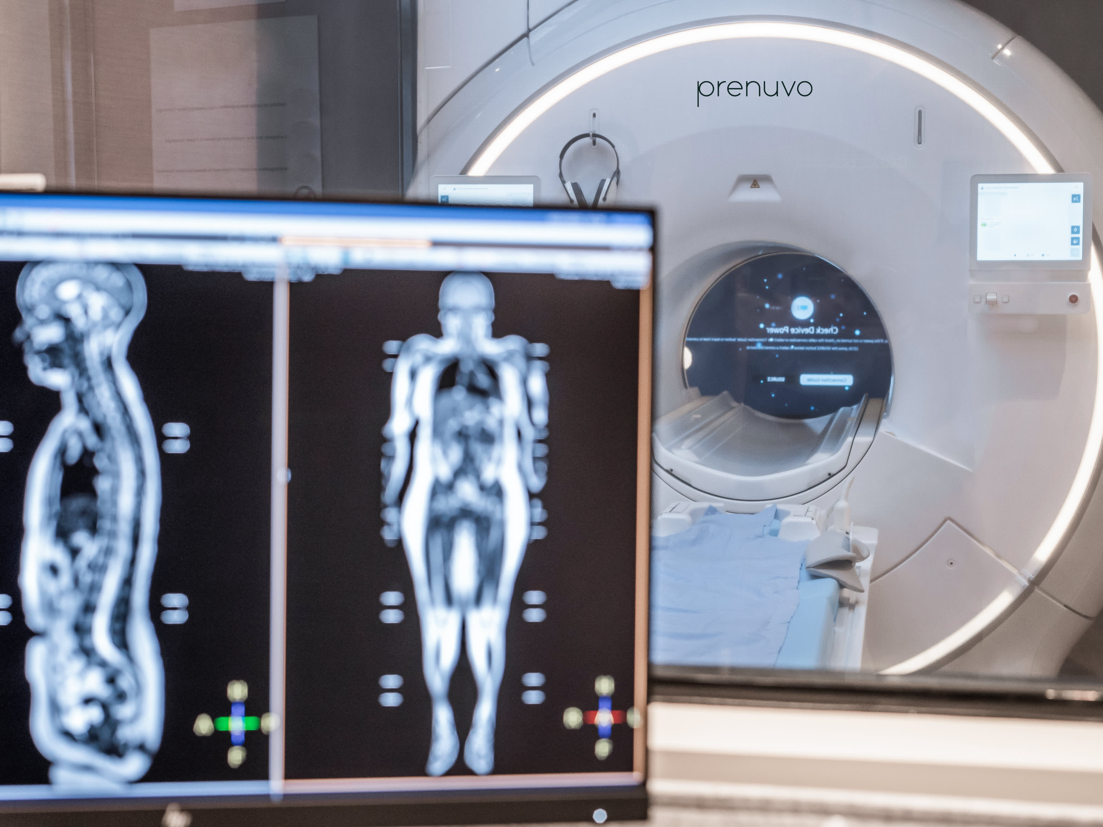
At its core, MRI relies on a powerful magnetic field, radio waves, and a sophisticated computer system. Unlike X-rays or CT scans, which use ionizing radiation, MRI utilizes the natural magnetic properties of atoms within the body, typically hydrogen atoms. When a patient is placed inside the MRI machine—a large tube surrounded by a strong magnet—the hydrogen atoms align with the magnetic field.
Capturing the Invisible
What sets MRI apart is its ability to capture high-resolution images of soft tissues, such as organs, muscles, and nerves. This capability is especially crucial for diagnosing conditions in the brain, spinal cord, joints, and abdomen, where traditional X-rays or CT scans may fall short in clarity or detail.
The process begins with the transmission of radio waves into the body, which temporarily disrupt the alignment of hydrogen atoms. As the atoms realign with the magnetic field, they emit signals that vary according to the type of tissue they are in. These signals are then detected by the MRI machine’s receiver coil and processed by a computer to construct detailed cross-sectional images.
Applications in Medicine
MRI scans are instrumental in diagnosing a wide range of conditions, including neurological disorders such as strokes, brain tumors, and multiple sclerosis. They are also invaluable in assessing joint and spinal injuries, detecting heart and vascular diseases, and evaluating the health of organs like the liver, kidneys, and reproductive system.
Beyond diagnosis, MRI plays a crucial role in planning and monitoring treatments. Surgeons use MRI images to precisely locate tumors and plan surgeries with minimal damage to surrounding healthy tissue. Oncologists monitor the response of tumors to chemotherapy or radiation therapy, adjusting treatment plans as needed based on real-time imaging results.
Advancements and Challenges
Recent advancements in MRI technology continue to push the boundaries of medical imaging. Techniques like functional MRI (fMRI) enable researchers to map brain activity and study cognitive functions in unprecedented detail. Diffusion tensor imaging (DTI) tracks the movement of water molecules in tissues, providing insights into the connectivity of nerve fibers in the brain and spinal cord.
However, MRI is not without its challenges. Patients with certain medical implants or metallic objects in their bodies may not be eligible for MRI scans due to safety concerns related to the strong magnetic field. Moreover, the cost and availability of MRI machines can limit access in some regions, posing disparities in healthcare delivery.
Looking Ahead
As researchers refine techniques and improve accessibility, the future of MRI holds promise for even greater advancements. Emerging technologies aim to enhance image resolution, shorten scan times, and reduce the need for contrast agents—all while making MRI more accessible to underserved populations worldwide.
In conclusion, Magnetic Resonance Imaging represents a pinnacle of medical imaging technology, offering unparalleled insights into the human body’s inner workings. From routine check-ups to critical diagnoses, MRI scans continue to shape the landscape of modern healthcare, paving the way for more precise, personalized, and effective medical treatments.
